













CT chest: second opinion
Tap an underlined word for its meaning
Clinical question
Lung cancer screening, first scan. Former smoker, 40 pack-years, stopped 6 years ago. Second opinion requested on the original report.
Technique
Low-dose CT of the chest without contrast, from the top of the lungs to the adrenal glands. 1.25 mm images in lung and soft-tissue windows, with coronal and sagittal views.
Comparison
No earlier chest CT available. The original report of this scan (21 Apr 2026) was reviewed.
Findings
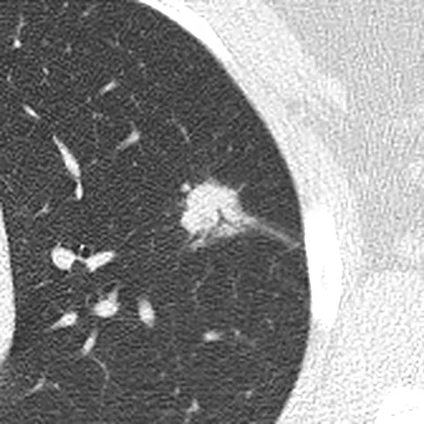
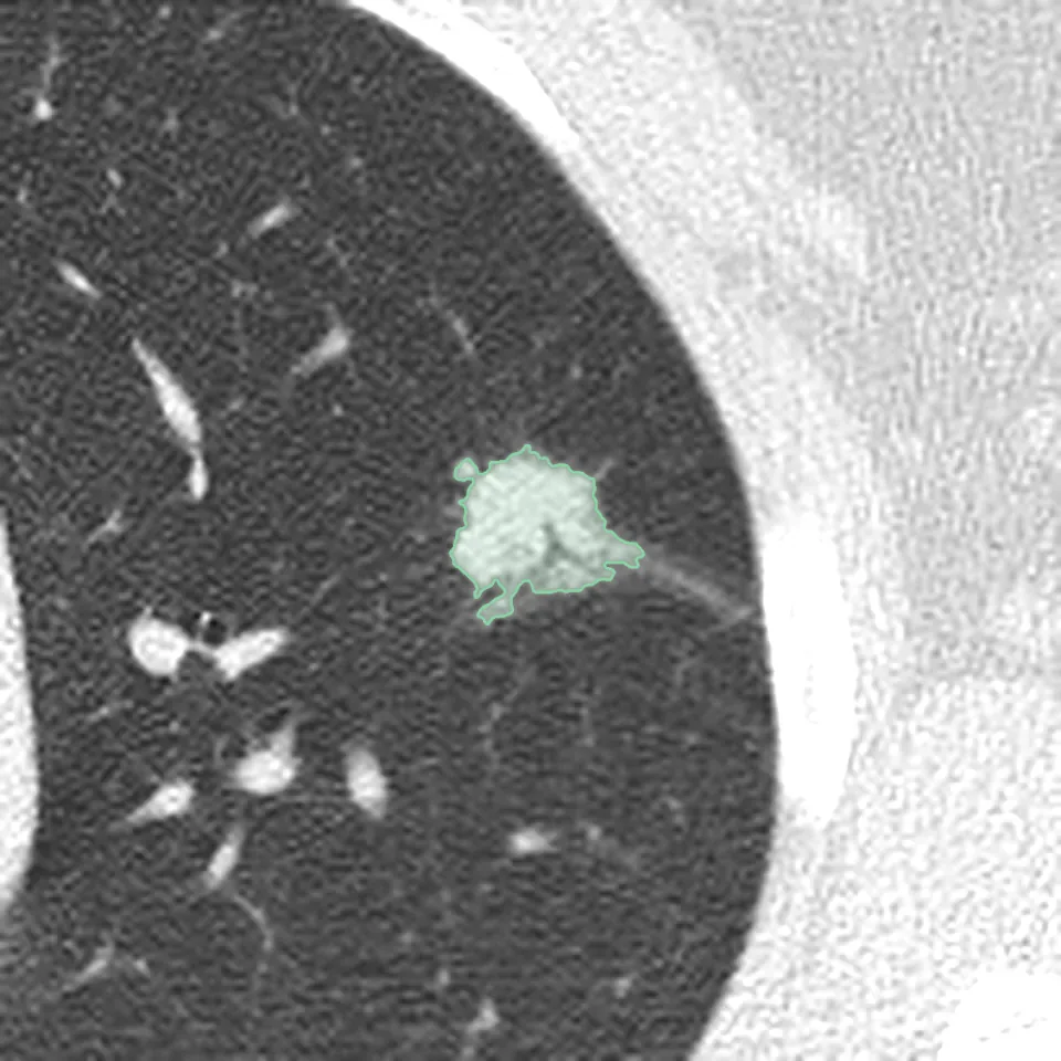
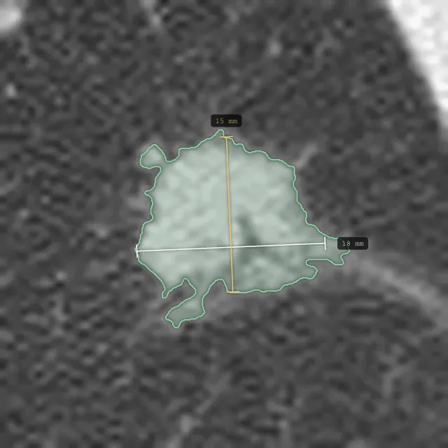
- Lungs. Solid nodule in the left upper lobe, 18 × 15 mm (series 2, image 190), with spiky edges (spiculated) and a thin strand pulling on the nearby lining of the lung (pleural tag).
- Lungs. Mild emphysema in both upper lobes. No other nodules.
- Airways. The windpipe and main airways are clear.
- Lymph nodes. No enlarged lymph nodes in the chest or at the roots of the lungs.
- Pleura. No fluid around the lungs (no pleural effusion).
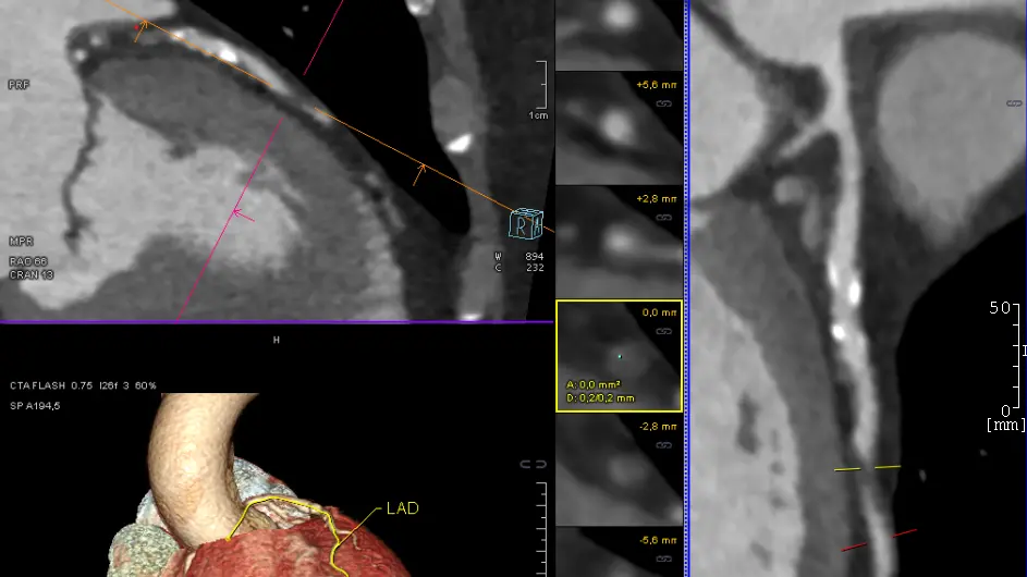
- Heart and vessels. Normal heart size. Mild calcium in the heart arteries (coronary calcification).
- Upper abdomen and bones. Nothing abnormal in the upper abdomen in view. Mild wear of the thoracic spine.
Conclusion
- A single 18 mm nodule in the left upper lobe with spiky edges. Its size and shape need prompt work-up (Lung-RADS 4X).
- No sign of spread to the lymph nodes or the lining of the lung.
- Mild emphysema and mild coronary calcification, both common in former smokers.
Compared with your original report
Agrees with your original report, which describes the same 18 mm nodule in the left upper lobe and advises further evaluation. This review adds three things: a risk category (Lung-RADS 4X, because of the spiky edge and the strand to the pleura), a clear next step with a time frame (PET-CT within two weeks), and the nodule outlined and measured on your images.
Recommendations
- PET-CT within two weeks to see how active the nodule is.
- Review by the lung team (thoracic multidisciplinary meeting) once the PET-CT is back.
- If the PET-CT is unclear, a CT-guided biopsy can be considered.
- Discuss the coronary calcium with your GP as part of a heart-health check.
Writing a plain-language explanation of your report. This takes a few seconds.
One spot in your upper left lung needs a prompt check with a PET-CT.
This low-dose CT looked at your chest. It found one 18 mm spot with spiky edges in the upper part of your left lung, which the report says needs prompt work-up. There is no sign of spread to the lymph nodes or the lining of the lung. It also found mild emphysema and mild calcium in the heart arteries.
What the report found
Spot in the upper left lung
A solid spot of 18 by 15 mm sits in the upper part of the left lung. Its edges are spiky, and a thin strand pulls on the nearby lining of the lung. The report says its size and shape need prompt work-up.
In the words of Dr. Sarah BennettSolid nodule in the left upper lobe, 18 × 15 mm (series 2, image 190), with spiky edges (spiculated) and a thin strand pulling on the nearby lining of the lung (pleural tag).
Mild emphysema
The report describes mild emphysema in the upper part of both lungs, and no other spots.
In the words of Dr. Sarah BennettMild emphysema in both upper lobes. No other nodules.
Calcium in the heart arteries
The heart is a normal size. The report sees mild calcium in the heart arteries and suggests talking about it with your GP as part of a heart-health check.
In the words of Dr. Sarah BennettNormal heart size. Mild calcium in the heart arteries (coronary calcification).
Mild wear of the spine
The report describes mild wear of the spine in the chest. The part of the upper belly in view looks normal.
In the words of Dr. Sarah BennettNothing abnormal in the upper abdomen in view. Mild wear of the thoracic spine.
- Windpipe and airways
The windpipe and the main airways are clear.
- Lymph nodes
No enlarged lymph nodes in the chest or where the lungs join the middle of the chest.
- Lining of the lungs
There is no fluid around the lungs.
What the report recommends
A PET-CT within two weeks, to see how active the spot is.
A review by the lung team once the PET-CT result is back.
If the PET-CT is unclear, a CT-guided biopsy can be considered.
Talking with your GP about the calcium in the heart arteries, as part of a heart-health check.
Images from your specialist
Questions you could ask
Tap one to put it in your chat with your specialist.
The explanations on this page are written by AI from the report and can contain mistakes. Where they differ from the report, the report is what counts.